Is lamina dura radiolucent
 Olivia House
 Radiographic signs of endodontic disease that are associated with the tissues around tooth roots include: Increased width of the apical radiolucent periodontal ligament space. Loss of the radiopaque lamina dura at the apex or other portals of exit such as lateral canals.
What does the lamina dura in the radiograph represent?
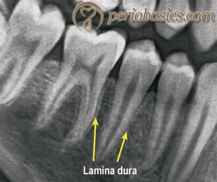
Lamina dura (LD) is a radiographic landmark viewed largely on periapical radiographs (PR). The terminology LD (or alveolus) is applied to the thin layer of dense cortical bone, which lines the roots of sound teeth. Presence of LD is an indication of the health of the teeth.
Is PDL radiopaque?
In radiographs, the PDL appears as the periodontal space of about 0.4 to 1.5 mm. It appears as a radiolucent area which is located between the radiopaque lamina dura of the alveolar bone proper and the radiopaque cementum.
What forms the lamina dura?
The alveolar process includes a region of compact bone that is adjacent to the periodontal ligament (PDL). This is called the lamina dura when it is viewed on radiographs. It is the lamina dura which is attached to the cementum from the roots by the periodontal ligament. It is uniformly lighter.What causes thickened lamina dura?
Lamina Dura Changes in Apical Periodontitis- caused by infection of the pulp canal system has been termed apical periodontitis, apical granuloma/cyst, periapical osteitis, periradicular periodontitis, among other terms.
When is lamina dura lost?
It is usually considered that the loss of the lamina dura is pathognomonic of hyperparathyroidism although some degree of loss may be apparent in osteomalacia and in Paget’s disease.
Which term is also used to refer to the lamina dura?
: the thin hard layer of bone that lines the socket of a tooth and that appears as a dense white line in radiography. — called also cribriform plate.
What is PDL widening?
A vertical bone defect develops when bone loss progresses down the root of the teeth in association with a deep periodontal pocket. In its early stage, this phenomenon appears as abnormal PDL widening (Fig. 3).What bone forms most of the lamina dura?
Alveolar bone proper appears on a radiograph as a thick radiopaque line adjacent to the alveolar socket, termed the lamina dura. The alveolar bone proper provides the attachment site for Sharpey fibers from the PDL.
Why is it called the alveolar ridge?It is so-named because the small jaw ridges are actually the edges of the cavity sockets, or alveoli, that house the roots of teeth. Although subtle and somewhat difficult to discern, they can be felt as an irregular and bumpy surface by the tongue tracing the hard palate near the inner base of teeth.
Article first time published onWhat is the function of the alveolar process?
The alveolar process is the thick ridge of bone in the jaw that holds the dental alveoli, or tooth sockets. The dental alveoli hold the roots of the teeth in place, and in case of a dental implant, the alveolar process holds implant hardware in place.
What is the alveolar process made of?
The alveolar process is the lining of the tooth socket and also known as the alveolus. While the alveolar process is made from compact bone, it can also be called the cribriform plate because it contains holes where Volkmann canals pass from the alveolar bone into the PDL.
Is the alveolar crest radiolucent or radiopaque?
The alveolar crest is the gingival margin of the alveolar process and extends between and around the tooth root(s). The crest is covered by a thin layer of cortical bone, appearing radiopaque radiographically (Figure 7).
Is the alveolar crest radiopaque?
In a normal and healthy periodontium, the alveolar crest is a thin, continuous radiopaque line that is continuous with the lamina dura (Figure 11.1).
Is PDL radiolucent or radiopaque?
Periodontal Ligament Space Because the PDL is composed primarily of collagen, it appears as a radiolucent space between the tooth root and the lamina dura.
Which cells lay down the alveolar bone?
Two major types of cells participate in the process—osteoblasts and osteoclasts. Osteoblasts in the alveolar bone originate directly from the dental mesenchyme (intramembranous ossification).
What is cause Hypercementosis?
Hypercementosis is excessive deposition of cementum on the tooth roots. In most cases, its cause is unknown. Occasionally, it appears on a supraerupted tooth after the loss of an opposing tooth. Another cause of hypercementosis is inflammation, usually resulting from rarefying or sclerosing osteitis.
What is the main cause of periodontal disease?
Periodontal (gum) disease is an infection of the tissues that hold your teeth in place. It’s typically caused by poor brushing and flossing habits that allow plaque—a sticky film of bacteria—to build up on the teeth and harden.
Where would the dental radiographer find the lamina dura in a dental image?
The lamina dura appears as a radiopaque line between the periodontal ligament and the alveolar socket.
What is the difference between radiopaque and radiolucent?
Radiolucent – Refers to structures that are less dense and permit the x-ray beam to pass through them. … Radiopaque – Refers to structures that are dense and resist the passage of x-rays. Radiopaque structures appear light or white in a radiographic image.
What is attached gingiva?
Attached gingiva – This tissue is adjacent to the free gingiva and is keratinized and firmly attached to the bone structure. It can range from 3-12 mm in height. Free gingiva – This tissue is not attached and forms a collar around the tooth.
What does periapical mean?
adjective. encompassing or surrounding the tip of the root of a tooth.
What are Sharpey's Fibres?
Sharpey’s fibres (bone fibres, or perforating fibres) are a matrix of connective tissue consisting of bundles of strong predominantly type I collagen fibres connecting periosteum to bone. … In the spine, similar fibres join the intervertebral disc to the adjacent vertebrae.
Which bones hold teeth?
The alveolar bone. It forms the tooth socket and provides the tooth with support. The fleshy tissue between the tooth and the tooth socket. It holds the tooth in place.
Which bones are just inferior to the Glabella?
A horizontal plate that forms the roof of the nasal cavity and closes the anterior part of the base of the cranium. The nasal bones lie directly inferior to the glabella. They from the bridge of the nose and the dome over the superior portion of the nasal cavity.
How does alveolar bone develop?
The alveolar bone begins to first form by an intramembranous ossification with in the ectomesenchyme surrounding the developing tooth. This first formed bone is called as woven bone is less organized and is replaced with more organized lamellar one. When a deciduous tooth is shed, its alveolar bone is resorbed.
Where is the PDL space?
Because the PDL is composed primarily of collagen, it appears as a radiolucent space between the tooth root and the lamina dura. This space begins at the alveolar crest, extends around the portions of the tooth roots within the alveolus, and returns to the alveolar crest on the opposite side of the tooth (Fig. 8-9).
Why is a sound called alveolar?
Alveolar consonants are consonant sounds that are produced with the tongue close to or touching the ridge behind the teeth on the roof of the mouth. The name comes from alveoli – the sockets of the teeth. … Alveolar consonants exist in many languages, including Spanish, Italian, French and German.
What is the function of alveolar ridge?
1 : the bony ridge or raised thickened border of the upper or lower jaw that contains the sockets of the teeth : alveolar process It is common for many of the teeth to be displaced from the alveolar ridge into the palate.
What does alveolar ridge mean?
The alveolar ridge is a small protuberance just behind the upper front teeth that can easily be felt with the tongue. The major part of the roof of the mouth is formed by the hard palate in the front, and the soft palate or velum at…
Does the alveolar process support the upper or lower teeth?
Alveolar Process: What You Should Know Your alveolar process (also known as the alveolar bone) is the structure that holds the roots of your teeth in place. You have an alveolar process made of thick bone for both your top and bottom rows of teeth.