What does C-arm machine do
 Natalie Ross
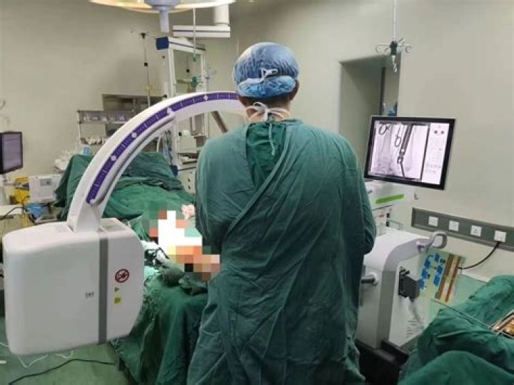
 In brief, a C-arm machine is a piece of medical imaging equipment that operates on the basic principle of X-ray technology. This fluoroscopy device is used to visualise patients’ anatomy in the operating room during surgery.
How does a C-arm work?
A c-arm uses an x-ray source (generator) and a flat-panel detector or image intensifier depending on your model. The C-shaped arm allows flexibility in movement so the x-rays of the patients can be viewed from all angles. The generator produces x-rays that infiltrate the patient’s body that is being scanned.
What is the difference between C arm and O arm?
O-arm navigation system use is shorter in radiation time and larger in radiation exposure than C-arm fluoroscopy navigation system. However, the amount of the radiation exposure per unit time in O-arm navigation system is larger than in C-arm fluoroscopy navigation system.
How much radiation do you get from C arm?
With ionising radiation produced by a standard C-arm, this is roughly equal to equivalent doses of 0.113 μSv per lateral image and 0.043 μSv per PA image 16.Where are C arms used?
C-Arm systems are used in the places where greater flexibility is required. They are being used in Orthopedic procedures, cardiac and angiography studies, and in therapeutic studies including stents and line placements.
How far from C-arm is safe?
Adjust distance. Your patient’s exposure to radiation increases exponentially by how close the patient is to the x-ray tube. Try to position your patient as far as possible from the tube. Ideally, your patient should be 12-15 inches away.
How do I unplug my C-arm?
The connector has a metal collar on the outside that slides back and releases the cable from the port. We often see cables that have been pulled from behind the connector, resulting in stress on both the housing and the wires, causing them to separate. Always unplug these cables by grabbing around the metal collar.
Where do you wear a radiation badge?
The Whole Body Radiation Badge is typically worn on the shirt, facing front. It should not be blocked or covered. If you are required to wear a lead apron for any reason, the badge should be worn on the collar outside the lead apron.Does radiation stay in the room?
The radiation stays in the body for anywhere from a few minutes to a few days. Most people receive radiation therapy for just a few minutes. Sometimes, people receive internal radiation therapy for more time. If so, they stay in a private room to limit other people’s exposure to the radiation.
How much does an O-arm cost?The O-arm integrates well with Stealth Navigation and costs A$850,000. Figures of the different 3D-fluoroscopy imaging systems are provided in Figure 1.
Article first time published onIs an O-arm a CT scanner?
The O-arm is a portable imaging device with a C-shaped arm that fits over the surgical table to take traditional two-dimensional X-ray images. During surgery it works like a CT scanner to take three-dimensional images in real time.
What is an O-arm in surgery?
The O-armTM system is an intraoperative 2D/3D imaging system that is designed to meet the workflow demands of the surgical environment. It can be used in variety of procedures including spine, cranial, and orthopedics.
Who invented the C-arm?
1955 Prior to 1955, X-ray systems were unable to change direction. Philips therefore developed the first C-arm – an X-ray system in the form of a half moon.
Who can operate a fluoroscopy machine?
Some state radiation safety regulations require fluoroscopic equipment operators to obtain special permits to perform fluoroscopy. For example, in the state of California, a radiologic technologist or a physician assistant must hold a California fluoroscopic permit to participate in fluoroscopic examinations [5,6].
Where is the image intensifier on a C-arm?
Most systems arranged as c-arms can have the image intensifier positioned above or below the patient (with the X-ray tube below or above respectively), although some static in room systems may have fixed orientations.
How do you use fluoroscopy?
During a fluoroscopy procedure, an X-ray beam is passed through the body. The image is transmitted to a monitor so the movement of a body part or of an instrument or contrast agent (“X-ray dye”) through the body can be seen in detail.
What is the purpose of the image intensifier?
An image intensifier or image intensifier tube is a vacuum tube device for increasing the intensity of available light in an optical system to allow use under low-light conditions, such as at night, to facilitate visual imaging of low-light processes, such as fluorescence of materials in X-rays or gamma rays (X-ray …
Why do you need to wear a valid radiation monitoring badge?
Note: If you are issued a badge, you must wear it whenever you are working near radiation. These badges provide legal records of accumulated radiation exposure for a lifetime; therefore, it is imperative that they are used when issued.
How far does scatter radiation travel?
A general rule of thumb is that the amount of scatter radiation at 1 meter (m) from the side of the patient will be 0.1% of the intensity of the primary x-ray beam.
How much radiation do you get from fluoroscopy?
Getting a fluoroscopic procedure exposes a patient to as much radiation as 250 to 3,500 chest X-rays. For perspective, a person gets the equivalent of one chest X-ray from normal background radiation in about two and a half days.
What should you not do during radiation?
Spicy Foods – Radiation often causes nausea, loose stools, or constipation. Spicy foods can further irritate the stomach and the rectum and cause discomfort. Raw Fish/Shellfish – Radiation therapy kills healthy cells in addition to cancerous cells, which could reduce the strength of your immune system.
Do tumors grow back after radiation?
Normal cells close to the cancer can also become damaged by radiation, but most recover and go back to working normally. If radiotherapy doesn’t kill all of the cancer cells, they will regrow at some point in the future.
What can I expect after my first radiation treatment?
The most common early side effects are fatigue (feeling tired) and skin changes. Other early side effects usually are related to the area being treated, such as hair loss and mouth problems when radiation treatment is given to this area. Late side effects can take months or even years to develop.
How often should radiation badges be changed?
Badges are exchanged quarterly. You should expect to receive your new badges a day or two before the start of each calendar quarter.
Why do radiation workers wear a film badge?
A film badge dosimeter monitors your radiation exposure to prevent you from exposure to over 10% of the allowable radiation limit (ALARA). The radiation badge measures the amount of radiation your body is exposed to so that you do not absorb large amounts of ionizing radiation.
What are the three key factors for limiting exposure to radiation?
- Time. Radiation exposure can be accumulated over the time of exposure. …
- Distance. A greater distance from the radiation source can reduce radiation exposure. …
- Shielding.
What is surgical imaging?
Imaging i.n.surgery. is used for diagnosis, planning, intraoperative navigation and post-operative evaluation, DIgItal medical imaging modalities mclude computed tomography (CT), magnetic resonance Imaging (MRI), MR therapy (MRT), fluoroscopy and ultrasound.
What is a Jackson table?
The Jackson Table (Fig. 1) method encompasses sliding the patient from a cart onto the table with appropriate padding placed while the patient is strapped securely on the table. The carbon fiber table frame is placed over the patient, and the patient-table construct is sandwiched together.
What is Stealth navigation?
Stealth technology guides surgical instruments with the potential for greater accuracy and precise sinus surgery. … Using Stealth technology, our team of ENT specialists are able to guide an endoscope inserted into the nasal passage, meaning that an open incision is rarely necessary.
Is the O-arm cone beam?
Purpose: The O-arm is a cone beam imaging system designed primarily to support orthopedic surgery as well as for image-guided and vascular surgery. Using a gantry that can be opened or closed, the O-arm can function as a 2D fluoroscopy device or collect 3D volumetric imaging data like a CT system.
What is AC arm radiology?
A C-arm is an imaging scanner intensifier. The name derives from the C-shaped arm used to connect the x-ray source and x-ray detector to one another. C-arms have radiographic capabilities, though they are used primarily for fluoroscopic intraoperative imaging during surgical, orthopedic and emergency care procedures.