Where is the septal nucleus
 Christopher Green
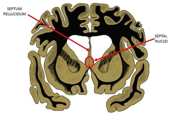
 The septal nuclei are present in most vertebrates, and in primates they are located medially in the cerebral hemispheres inferior to the rostrum of the corpus callosum and anterior to the third ventricle (Fig. 1; Mark et al., 1994).
What are the septal nuclei?
The septal nuclei are composed of medium-size neurons which are classified into dorsal, ventral, medial, and caudal groups. The septal nuclei receive reciprocal connections from the olfactory bulb, hippocampus, amygdala, hypothalamus, midbrain, habenula, cingulate gyrus, and thalamus.
What part of the brain is the septum?
Where is the septum? The term septum, when used in reference to the brain (it is a common anatomical term used to refer to a partition), indicates a subcortical structure in the forebrain that is found near the midline of the brain.
Where is medial septal nucleus?
The triangular and medial septal nuclei are in the midline. The anterior medial septal nucleus blends in with the vertical limb of the diagonal band of Broca just lateral to the midline. A large lateral septal nucleus, situated on either side of the midline nuclei, extends throughout the entire anteroposterior extent.What is the septal area associated with?
The septal area is a subcortical region that has strong projections to emotion-generating areas and has a key role in feelings of social connectedness and bonding. In rats, oxytocin binding in the septal area has been associated with maternal behaviors that promote kinship bonds (Francis, Champagne, & Meaney, 2000).
What does the nucleus accumbens do?
Introduction: The nucleus accumbens is considered as the neural interface between motivation and action, playing a key role on feeding, sexual, reward, stress-related, drug self-administration behaviors, etc.
What does the septum in the brain do?
Function. The septum is considered a part of the limbic system, mediating the connection between the cortex and subcortical limbic nuclei. The septum projects fibres to the hypothalamus, hippocampus, amygdala, reticular formation and olfactory cortical areas, suggesting a role in limbic regulation.
What is brain fornix?
The fornix is a white matter bundle located in the mesial aspect of the cerebral hemispheres, which connects various nodes of a limbic circuitry and is believed to play a key role in cognition and episodic memory recall.What is the Septo hippocampal system?
The septo–hippocampal pathway adjusts CA1 network excitability to different behavioral states and is crucially involved in theta rhythmogenesis. … Neurons of these three classes project to glutamatergic pyramidal neurons and different subsets of GABAergic neurons in the hippocampal CA1 region.
What is medial septum?The medial septal nucleus (MS) is one of the septal nuclei. Neurons in this nucleus give rise to the bulk of efferents from the septal nuclei. A major projection from the medial septal nucleus terminates in the hippocampal formation. It plays a role in the generation of theta waves in the hippocampus.
Article first time published onWhat does the septum pellucidum separate?
The septum pellucidum (meaning translucent wall in Latin – SP), also known as the ventricle of Sylvius, is a thin, triangular double membrane separating the frontal horns of the right and left lateral ventricles of the brain.
What is absent septum pellucidum?
The absence of the septum pellucidum is a rare condition that affects the structure of the brain. Specifically, a thin membrane called the septum pellucidum is missing from its normal position in the middle of the brain.
What is a basal nucleus?
The basal ganglia, or basal nuclei, are a group of subcortical structures found deep within the white matter of the brain. They form a part of the extrapyramidal motor system and work in tandem with the pyramidal and limbic systems.
Where is the septal wall?
The septum is the muscular wall that separates the left and right side of the heart. Problems occur when the septum between the heart’s lower chambers, or ventricles, is thickened.
Why is the septum so important?
When a person has a VSD, there’s an opening in the septum between the left ventricle and right ventricle. This part of the septum is called the ventricular septum. This hole allows blood from the left ventricle to go back into the right ventricle instead of out of the heart through the aorta.
Why is the septum pellucidum important?
The septum pellucidum is a thin, laminated translucent vertical membrane in the midline of the brain separating the anterior horns of the right and left ventricles. … This is an important normal structure to identify in the sonographic assessment of the fetal brain.
What is the pleasure center of the brain?
Located near the center of the brain, the nucleus accumbens is connected, by intermingled populations of cells, to many other brain structures having roles in pleasure seeking and drug addiction.
Where in the brain is the nucleus accumbens?
The nucleus accumbens is found in an area of the brain called the basal forebrain. There is a nucleus accumbens in each cerebral hemisphere; it is situated between the caudate and putamen. The nucleus accumbens is considered part of the basal ganglia and also is the main component of the ventral striatum.
Where is dopamine produced in the brain?
Dopamine producing neurons are located in the midbrain nuclei; mainly ventral tegmental area (VTA) and substantia nigra pars compacta (Poulin et al., 2018). Noradrenergic nuclei are located in pons and medulla.
What is limbic system?
The limbic system is a set of structures of the brain. … There are several important structures within the limbic system: the amygdala, hippocampus, thalamus, hypothalamus, basal ganglia, and cingulate gyrus.
Where is the 3rd ventricle?
The third ventricle is a narrow, funnel-shaped structure that lies in the center of the brain. It lies below the corpus callosum and body of the lateral ventricles, between the two thalami and walls of hypothalamus, and above the pituitary and midbrain (Fig.
Where is the cortical?
The cerebral cortex is a sheet of neural tissue that is outermost to the cerebrum of the mammalian brain. It has up to six layers of nerve cells. It is covered by the meninges and often referred to as gray matter.
Where does fornix end?
The anterior fibers (precommissural fornix) end at the septal nuclei of the basal forebrain and nucleus accumbens of each half of the brain.
What is the nucleus basalis of meynert?
The nucleus basalis of Meynert (nbM) was first described at the end of the 19th century and named after its discoverer, Theodor Meynert. The nbM contains a large population of cholinergic neurons that project their axons to the entire cortical mantle, the olfactory tubercle, and the amygdala.
What is thalamus function?
Generally, the thalamus acts as a relay station filtering information between the brain and body. Except for olfaction, every sensory system has a thalamic nucleus that receives, processes, and sends information to an associated cortical area.
Where is the septum linguae?
The lingual septum consists of a vertical layer of fibrous tissue, extending throughout the entire length of the median plane of the tongue, though not quite reaching the dorsum.
Is septum pellucidum paired?
The lamina of septum pellucidum is a paired layer forming the septum pellucidum.
Is the brain a solid organ that lacks cavities?
The brain is not a solid organ. Instead, there are fluid-filled cavities within the brain called ventricles.
Can people be born without a septum?
Absence of the septum pellucidum can be caused by multiple factors depending on the underlying condition. Some people are born with or develop hydrocephalus , or a fluid buildup in the brain. If this condition goes untreated, the excess fluid can disintegrate the septum pellucidum.
Does the septum pellucidum secrete cerebrospinal fluid?
During early development, the septum pellucidum is formed by the thinned walls of the 2 cerebral hemispheres and contains a fluid-filled cavity, named the cavum, which may persist. The ventricular system of the human brain. … Cerebrospinal fluid (CSF) is secreted by the choroid plexuses, filling the ventricular system.
Which ventricles are divided by the septum pellucidum?
The septum pellucidum (Latin for “translucent wall”) is a thin, triangular, vertical double membrane separating the anterior horns of the left and right lateral ventricles of the brain. It runs as a sheet from the corpus callosum down to the fornix.